
Case Report: Retinal nerve fiber layer and ganglion cell thinning on spectral domain optical coherence tomography following multiple strokes
DOI:
https://doi.org/10.15353/cjo.v84i3.1556Keywords:
RNFL, ganglion cell, optical coherence tomography, quadrantanopia, strokeAbstract
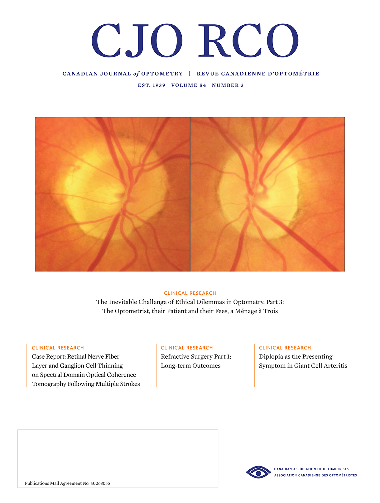
A 62-year-old Caucasian male presented for a routine exam with a history of a significant stroke suffered 5 years prior resulting in temporary aphasia and persistent visual field loss. At presentation, best correctable acuities were 20/20+ OU through mild hyperopic astigmatic correction. Visual field testing with frequency doubling technology revealed an incongruous left superior quandrantanopia. Anterior segments demonstrated mild bilateral mixed blepharitis but were otherwise unremarkable. Dilated fundus examination with spectral domain optical coherence tomography revealed moderate disc cupping and marked, symmetric inferior and temporal RNFL thinning, as well as diffuse bilateral ganglion cell thinning. This case demonstrates how OCT can be used as an objective test to detect retrograde neve fiber and ganglion cell loss secondary to post-chiasmal pathology.
Published
How to Cite
Issue
Section
License
Copyright (c) 2022 Nicholas A. Froumis, BSc, OD

This work is licensed under a Creative Commons Attribution-NonCommercial-NoDerivatives 4.0 International License.
