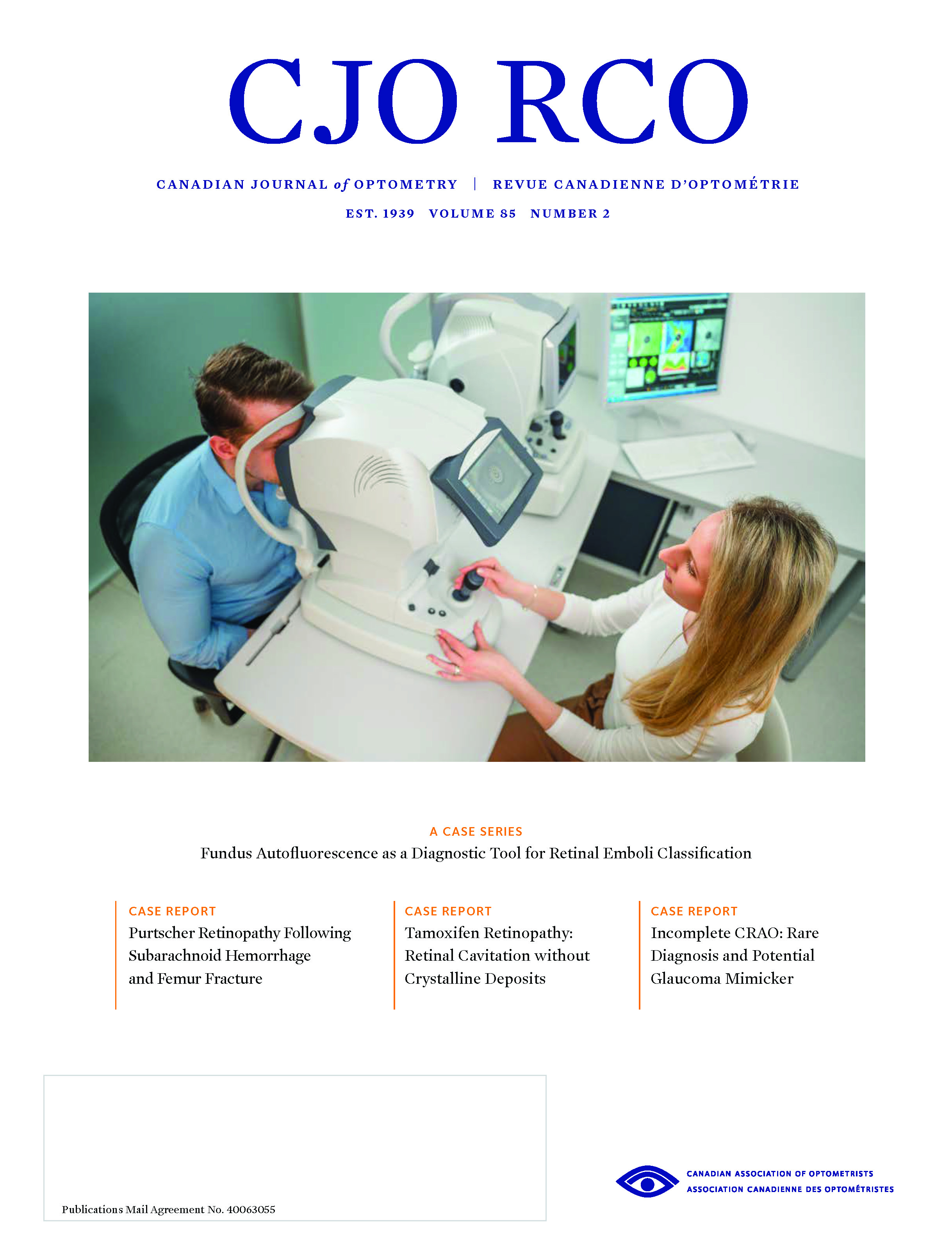
Fundus Autofluorescence as a Diagnostic Tool for Retinal Emboli Classification: A Case Series
Abstract
Fundus autofluorescence (FAF) is a relatively new imaging technique typically obtained with an enhanced fundus camera or scanning laser ophthalmoscope that is becoming more widely utilized in optometric practices. The images obtained can provide additional diagnostic information for a wide variety of retinal pathology. This case series highlights the benefit of using FAF to visualize retinal emboli. Specifically, the cases demonstrate how this technology can help identify embolus composition, allow better visualization of an embolus within the optic nerve, and differentiate an embolus from adjacent vascular sheathing.
References
Mitchell P, Wang JJ, Li W, Leeder SR, Smith W. Prevalence of asymptomatic retinal emboli in an Australian urban community. Stroke 1997;28(1):63-6. doi:10.1161/01.str.28.1.63
Cheung N, Teo K, Zhao W, et al. Prevalence and Associations of Retinal Emboli With Ethnicity, Stroke, and Renal Disease in a Multiethnic Asian Population: The Singapore Epidemiology of Eye Disease Study. JAMA Ophthalmol 2017;135(10):1023-1028. doi:10.1001/jamaophthalmol.2017.2972
Hollenhorst RW. Significance of bright plaques in the retinal arterioles. Trans Am Ophthalmol Soc 1961;59:252-73.
O'Donnell BA, Mitchell P. The clinical features and associations of retinal emboli. Aust N Z J Ophthalmol 1992;20(1):11-7. doi:10.1111/j.1442-9071.1992.tb00697.x
Bruno A, Russell PW, Jones WL, Austin JK, Weinstein ES, Steel SR. Concomitants of asymptomatic retinal cholesterol emboli. Stroke 1992;23(6):900-2. doi:10.1161/01.str.23.6.900
Wijman CA, Babikian VL, Matjucha IC. Monocular visual loss and platelet fibrin embolism to the retina. J Neurol Neurosurg Psychiatry 2000;68(3):386-7. doi:10.1136/jnnp.68.3.386
Bacquet JL, Sarov-Rivière M, Denier C, et al. Fundus autofluorescence in retinal artery occlusion: A more precise diagnosis. J Fr Ophthalmol 2017;40(8):648-53. doi:10.1016/j.jfo.2017.03.010
Sharma S, Pater JL, Lam M, Cruess AF. Can different types of retinal emboli be reliably differentiated from one another? An inter- and intraobserver agreement study. Can J Ophthalmol 1998;33(3):144-8
Wong TY, Larsen EK, Klein R, et al. Cardiovascular risk factors for retinal vein occlusion and arteriolar emboli: the Atherosclerosis Risk in Communities & Cardiovascular Health studies. Ophthalmology 2005;112(4):540-7. doi:10.1016/j.ophtha.2004.10.039
Klein R, Klein BE, Moss SE, Meuer SM. Retinal emboli and cardiovascular disease: the Beaver Dam Eye Study. Arch Ophthalmol 2003;121(10):1446-51. doi:10.1001/archopht.121.10.1446
Hoki SL, Varma R, Lai MY, Azen SP, Klein R; Los Angeles Latino Eye Study Group. Prevalence and associations of asymptomatic retinal emboli in Latinos: the Los Angeles Latino Eye Study (LALES). Am J Ophthalmol 2008;145(1):143-8. doi:10.1016/j.ajo.2007.08.030
Kleefeldt N, Bermond K, Tarau IS, et al. Quantitative Fundus Autofluorescence: Advanced Analysis Tools. Transl Vis Sci Technol 2020;9(8):2. Published 2020 Jul 1. doi:10.1167/tvst.9.8.2
Schmitz-Valckenberg S, Holz FG, Bird AC, Spaide RF. Fundus autofluorescence imaging: review and perspectives. Retina 2008;28(3):385-409. doi:10.1097/IAE.0b013e318164a907
Sato T, Mrejen S, Spaide RF. Multimodal imaging of optic disc drusen. Am J Ophthalmol 2013;156(2):275-82.e1. doi:10.1016/j.ajo.2013.03.039
Siddiqui AA, Paulus YM, Scott AW. Use of fundus autofluorescence to evaluate retinal artery occlusions. Retina 2014;34(12):2490-2491. doi:10.1097/IAE.0000000000000186
Munk MR, Mirza RG, Jampol LM. Imaging of a cilioretinal artery embolisation. Int J Mol Sci 2014;15(9):15734-40. Published 2014 Sep 4. doi:10.3390/ijms150915734
Rajesh B, Hussain R, Giridhar A. Autofluorescence and Infrared Fundus Imaging for Detection of Retinal Emboli and Unmasking Undiagnosed Systemic Abnormalities. J Ophthalmic Vis Res 2016;11(4):449-51. doi:10.4103/2008-322X.194149
McLeod SD, Emptage NP, Harris JK, et al. Retinal and artery occlusions preferred practice pattern. American Academy of Ophthalmology. 2016.
Riese N, Smart Y, Bailey M. Asymptomatic retinal emboli and current practice guidelines: a review [published online ahead of print, 2022 Feb 2]. Clin Exp Optom 2022;1-6. doi:10.1080/08164622.2022.2033600
Ahmmed AA, Carey PE, Steel DH, Sandinha T. Assessing Patients with Asymptomatic Retinal Emboli Detected at Retinal Screening. Ophthalmol Ther 2016;5(2):175-82. doi:10.1007/s40123-016-0055-5
Ahmed R, Khetpal V, Merin LM, Chomsky AS. Case Series: Retrospective Review of Incidental Retinal Emboli Found on Diabetic Retinopathy Screening: Is There a Benefit to Referral for Work-Up and Possible Management?. Clin Diabetes 2008;26(4):179. doi:10.2337/diaclin.26.4.179
McCullough HK, Reinert CG, Hynan LS, et al. Ocular findings as predictors of carotid artery occlusive disease: is carotid imaging justified?. J Vasc Surg 2004;40(2):279-86. doi:10.1016/j.jvs.2004.05.004
Bakri SJ, Luqman A, Pathik B, Chandrasekaran K. Is carotid ultrasound necessary in the evaluation of the asymptomatic Hollenhorst plaque?. Ophthalmology 2013;120(12):2747-8.e1. doi:10.1016/j.ophtha.2013.09.005
Hadley G, Earnshaw JJ, Stratton I, Sykes J, Scanlon PH. A potential pathway for managing diabetic patients with arterial emboli detected by retinal screening. Eur J Vasc Endovasc Surg 2011;42(2):153-7. doi:10.1016/j.ejvs.2011.04.031
Ramakrishna G, Malouf JF, Younge BR, Connolly HM, Miller FA. Calcific retinal embolism as an indicator of severe unrecognised cardiovascular disease. Heart. 2005;91(9):1154-7. doi:10.1136/hrt.2004.041814
Published
How to Cite
Issue
Section
License
Copyright (c) 2023 Nicole Auchter Riese, Yelena Smart, Tara Foltz, Drew Anderson

This work is licensed under a Creative Commons Attribution-NonCommercial-NoDerivatives 4.0 International License.


